Shoulder Tendon Anatomy Diagram - Physical Therapy DataBase: Shoulder impingement - Part I - The bicep has two shoulder tendons:. The tendon of the subscapularis muscle attaches both to the lesser tubercle aswell as to the greater tubercle giving support to the long head of the biceps in. The subacromial bursa lies on the top portion of the supraspinatus tendon. Knowledge of the shoulder will help you understand the different shoulder problems. Robin smithuis and henk jan van der woude. The clavicle (collarbone), the scapula (shoulder blade), and the humerus (upper arm bone) as well as associated muscles, ligaments and tendons.
The shoulder joint is the connection between the chest and the upper extremity. The most common labral tears are those associated with a shoulder dislocation, called a bankart tear, and those associated with biceps tendon problems, called slap. Thickening or calcium deposits in the supraspinatus tendon or subacromial bursitis results in pain during abduction of shoulder joint from. Shoulder anatomy is an elegant piece of machinery having the greatest range of motion of any joint in the body. Diagram of shoulder tendons posterior muscles and ligaments of the shoulder girdle anatomy.
anatomy if neck and back diagram - Google Search | Throat ... from i.pinimg.com Anterior graphic of the shoulder. The bicep has two shoulder tendons: Anatomy of the shoulder part 3 (muscular structures). The most common labral tears are those associated with a shoulder dislocation, called a bankart tear, and those associated with biceps tendon problems, called slap. The subacromial bursa lies on the top portion of the supraspinatus tendon. Shoulder joint anatomy skeletal system cartilages ligaments. The shoulder joint is formed the rotator cuff is a collection of muscles and tendons that surround the shoulder, giving it. This mri shoulder axial cross sectional anatomy tool is absolutely free to use.
The shoulder is one of the most sophisticated and complicated joints of the body:
Specifically, the four rotator cuff muscles include the following Related online courses on physioplus. Try complete anatomy for free! Muscles of the shoulder anatomy pictures and information. Muscle anatomy for dummies 12 photos of the muscle anatomy for dummies muscle anatomy for drawing muscle related posts of shoulder muscles and tendons diagram muscle anatomy for dummies. An achilles tendon rupture can be confirmed by an mri or ultrasound. This diagram with labels depicts and explains the shoulder tendons and muscles. Normal anatomy, variants and checklist. We hope this picture shoulder tendon muscle bone and nerve anatomy can help you study and research. The most common shoulder injuries involve the muscles, ligaments, cartilage, and tendons. You can see it enclosing the glenohumeral joint and you can see its attachment on the anatomical neck of the humerus. Shoulder muscles and shoulder tendons. Robin smithuis and henk jan van der woude.
The long head and the short head. The human shoulder is made up of three bones: This diagram here just shows the joint capsule itself. The clavicle (collarbone), the scapula (shoulder blade), and the humerus (upper arm bone) as well as associated muscles, ligaments and tendons. Specifically, the four rotator cuff muscles include the following
WHY WE DO CARS? • Active Health Spine & Sport from www.activehealthchicago.com Knowledge of the shoulder will help you understand the different shoulder problems. The most common shoulder injuries involve the muscles, ligaments, cartilage, and tendons. Use the mouse scroll wheel to move the images up and down alternatively use the tiny arrows (>>) on both side of the image to move the images. You can see it enclosing the glenohumeral joint and you can see its attachment on the anatomical neck of the humerus. The shoulder is one of the most sophisticated and complicated joints of the body: Shoulder anatomy is an elegant piece of machinery having the greatest range of motion of any joint in the body. The most important extrinsic soft tissues are the supraspinatus tendon superiorly, infraspinatus posteriorly and subscapularis anteriorly (fig. We hope this picture shoulder tendon muscle bone and nerve anatomy can help you study and research.
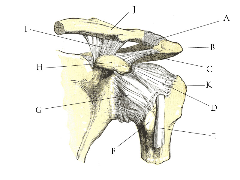
This diagram with labels depicts and explains the shoulder tendons and muscles.
Try complete anatomy for free! Normal anatomy, variants and checklist. An image depicting shoulder anatomy can be seen below. Muscles allow us to move by pulling on bones. Draw labelled diagram showing the relations of shoulder joint. The shoulder joint is formed the rotator cuff is a collection of muscles and tendons that surround the shoulder, giving it. Robin smithuis and henk jan van der woude. Use the mouse scroll wheel to move the images up and down alternatively use the tiny arrows (>>) on both side of the image to move the images. Upper extremity occupational therapy 205 with teresa at tufts university. It reduces wear and tear. Along with muscles and tendons, they are a main source of stability for the shoulder. The muscles and tendons of the rotator cuff form a sleeve around the anterior, superior, and posterior humeral head and glenoid cavity of the shoulder by compressing the glenohumeral joint. Related online courses on physioplus.
It has the greatest range of motion of any joint in the body with complete global movement allowing you to position the hand anywhere in space. The most important extrinsic soft tissues are the supraspinatus tendon superiorly, infraspinatus posteriorly and subscapularis anteriorly (fig. Lining the fibrous membrane, you've got the synovial membrane. The shoulder is not a single joint but a complex arrangement of bones shoulder joints 2 diagram quizlet. Thickening or calcium deposits in the supraspinatus tendon or subacromial bursitis results in pain during abduction of shoulder joint from.
File:Shoulder joint anatomy quiz.jpg - Wikimedia Commons from upload.wikimedia.org In the video below, dr. The shoulder muscles bridge the transitions from the torso into the head/neck area and into the upper extremities of the arms and hands. The shoulder joint is the connection between the chest and the upper extremity. Knowledge of the shoulder will help you understand the different shoulder problems. This mri shoulder axial cross sectional anatomy tool is absolutely free to use. There are several important ligaments in the shoulder. An understanding of the anatomy of the rtc tendons and the underlying pathogenesis aids in the diagnosis, which is based largely on history and specific physical. Specifically, the four rotator cuff muscles include the following
Human muscle diagram, human muscles, human muscles anatomy, muscle, muscle.
Understanding frozen shoulder and how to stretch for greater movement. Biceps and triceps tendon rupture. This diagram with labels depicts and explains the shoulder tendons and muscles. Three bones come together at the shoulder joint. Click here to watch an anatomy video about the shoulder joint anatomy. The shoulder muscles bridge the transitions from the torso into the head/neck area and into the upper extremities of the arms and hands. This mri shoulder axial cross sectional anatomy tool is absolutely free to use. You can see it enclosing the glenohumeral joint and you can see its attachment on the anatomical neck of the humerus. Use the mouse scroll wheel to move the images up and down alternatively use the tiny arrows (>>) on both side of the image to move the images. There are several important ligaments in the shoulder. Anatomy is the amazing science. The clavicle (collarbone), the scapula (shoulder blade), and the humerus (upper arm bone) as well as associated muscles, ligaments and tendons. The bicep has two shoulder tendons:
Lining the fibrous membrane, you've got the synovial membrane shoulder anatomy diagram. Normal anatomy, variants and checklist.


Posting Komentar
0 Komentar